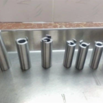
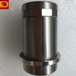
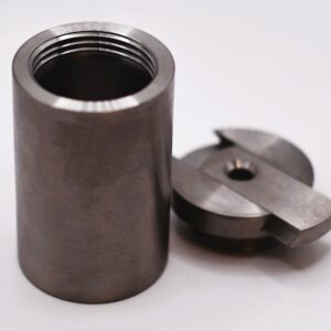
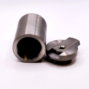
PET & SPECT Tungsten Alloy Vial Shields
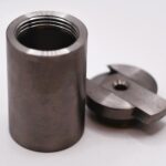
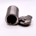
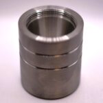
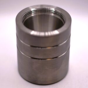
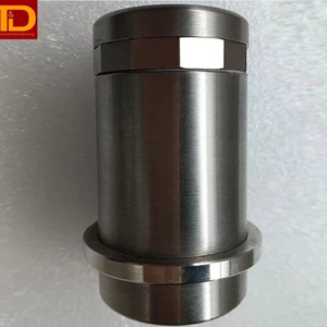
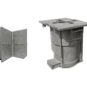
Tungsten Vial Shields for PET are designed to ensure maximum safety and precision in PET radiopharmaceutical applications. Made from tungsten, this shield offers unparalleled protection against radiation exposure, safeguarding both patients and healthcare professionals during imaging procedures. Its design ensures effortless handling and compatibility with standard PET vials, making it an essential addition to any medical facility seeking superior radiation shielding.
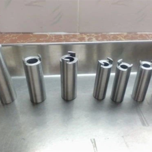
We offer custom Tungsten Vial Shields made according to your specifications and technical drawings. Our team works closely with you to manufacture a custom design that meets your specific requirements, whether it’s accommodating different vial sizes, optimizing shielding thickness, or incorporating additional features. Contact our team with any questions and for further details.
Tungsten Alloy Vial Shield for Positron Emission Tomography (PET)
Positron emission tomography (PET) is one of the nuclear medicine techniques for diagnosis. While X-rays reveal details on the structure of the body, PET provides insight into the chemical function of a specific organism. During PET imaging, FDG, a glucose-based radionuclide, is injected from a shielded syringe into the patient. As FDG moves throughout the body, it emits gamma radiation, detectable by a gamma camera. This process allows chemical activity within cells and organs to be seen. Anomalies in chemical activity may indicate the presence of tumors. PET scans are commonly employed to detect cancerous tumors as well as conditions affecting the brain and coronary arteries.
Applications for tungsten alloy shielding in PET include:
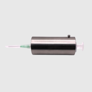
- PET Syringe Shielding
- Tungsten Vial Shielding
- Collimator for gamma camera
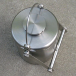
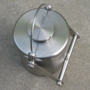
- Tungsten FDG transport container
Tungsten Alloy Vial Shield for Multi-Leaf Collimator
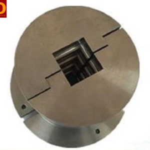
Radiotherapy destroys cancer cells by precisely targeting beams of radiation onto the tumor. These radiation beams require pinpoint accuracy to spare the surrounding healthy tissue from harm. This is achieved by using a multi-leaf collimator, composed of two rows of exceptionally thin tungsten alloy plates, which can be meticulously configured to match the exact dimensions of the tumor.

